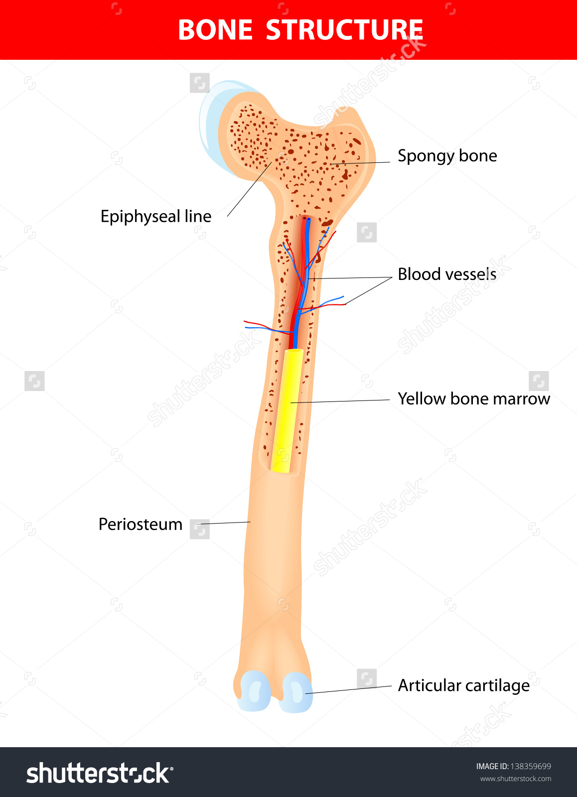
describe the femur with diagram
The X-rays of case 1. a Periprosthetic femoral fracture Vancouver C. 9 Images about The X-rays of case 1. a Periprosthetic femoral fracture Vancouver C : The Lower Limb | Boundless Anatomy and Physiology, The left panel shows the superficial pelvic and thigh muscles, the and also X-ray 2 (comminuted, displaced fracture). | Download Scientific Diagram.
The X-rays Of Case 1. A Periprosthetic Femoral Fracture Vancouver C
 www.researchgate.net
periprosthetic femoral initially distal
Anatomy 411 Quiz 2 Flashcards | Quizlet
 quizlet.com
lateral condyle medial tuberosity tibia quiz anatomy head malleolus fibula tibial quizlet gilroy pg
Fitting Of The TomoFix Medial Distal Femoral Plate (MDF; DePuy Synthes
 www.researchgate.net
plate distal femoral medial synthes tomofix depuy femur mdf
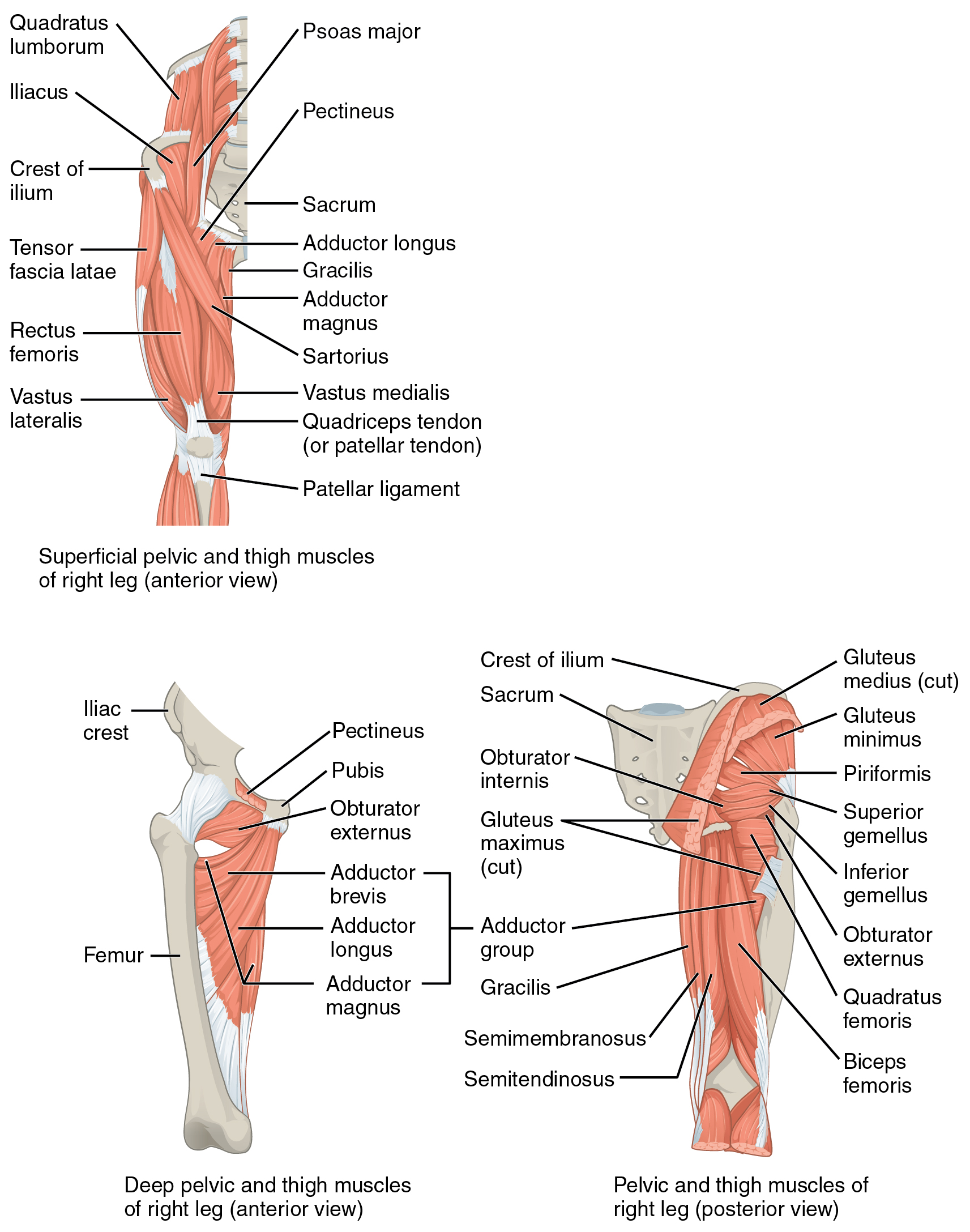
The Left Panel Shows The Superficial Pelvic And Thigh Muscles, The
 oerpub.github.io
muscles anatomy thigh hip femur pelvic lower move gluteal girdle appendicular limbs physiology leg muscle anterior posterior deep thighs hips
Diagram Demonstrating Measurement Of Glenoid Tilt. (Reprinted With
 www.researchgate.net
glenoid glenohumeral demonstrating reprinted
Skeletal System Long Bone Diagram / The Skeletal System - The
 neringuciuknews.blogspot.com
skeletal bones
X-ray 2 (comminuted, Displaced Fracture). | Download Scientific Diagram
 www.researchgate.net
fracture comminuted displaced
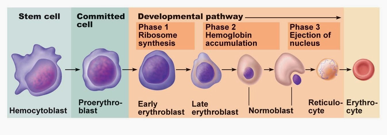
Erythropoiesis ~ ජීවක අරණ
 jeewakaarana.blogspot.com
erythropoiesis maturation
The Lower Limb | Boundless Anatomy And Physiology
 courses.lumenlearning.com
knee anatomy joint tibia femur patella physiology position articulation lower limb relative diagram boundless shows
Fracture comminuted displaced. Glenoid glenohumeral demonstrating reprinted. The x-rays of case 1. a periprosthetic femoral fracture vancouver c