ct temporal bone anatomy
How to Read a Head CT - Emergency Medicine | NewYork-Presbyterian. 9 Pictures about How to Read a Head CT - Emergency Medicine | NewYork-Presbyterian : Exported LEFT TEMPORAL FIBROUS DYSPLASIA, The Radiology Assistant : Temporal bone - Anatomy 2.0 and also Radiation Dose to the Lens Using Different Temporal Bone CT Scanning.
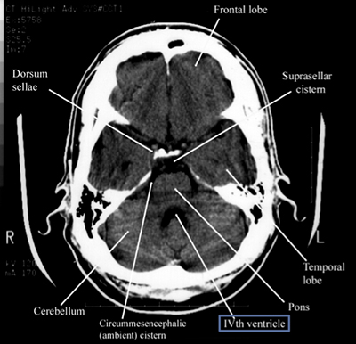
How To Read A Head CT - Emergency Medicine | NewYork-Presbyterian
 www.nyp.org
ct cistern head ventricles suprasellar read cisterns reading ivth emergency lateral
Fig 1. | Multisection CT Venography Of The Dural Sinuses And Cerebral
 www.ajnr.org
venography cerebral dural sinuses venogram angiography elimination multisection ctv ajnr
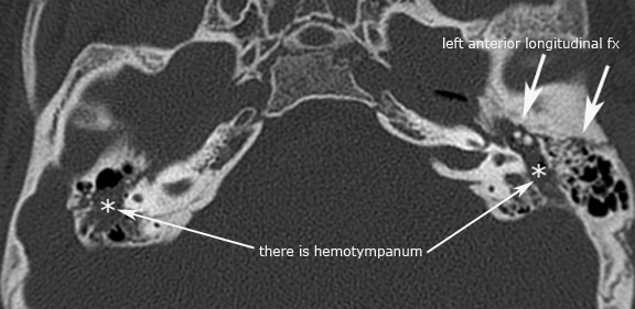
Longitudinal Temporal Bone Fracture | Image | Radiopaedia.org
 radiopaedia.org
fracture bone temporal longitudinal coronal radiopaedia ct mastoid contrast version non brain
Trauma
 uwmsk.org
bone fracture temporal longitudinal fractures trauma
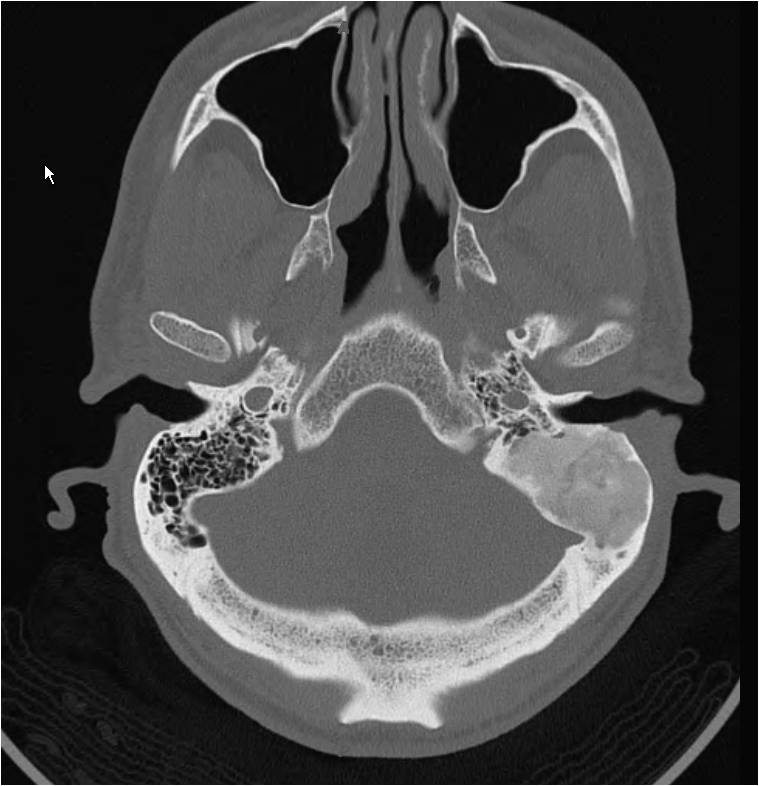
Skull Base Osteomyelitis | Image | Radiopaedia.org
 radiopaedia.org
osteomyelitis radiopaedia axial
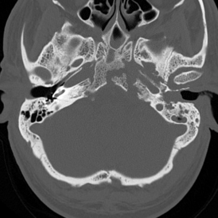
Radiation Dose To The Lens Using Different Temporal Bone CT Scanning
 www.ajnr.org
ct temporal bone head axial dose radiation scanning protocols lens different using ajnr fig
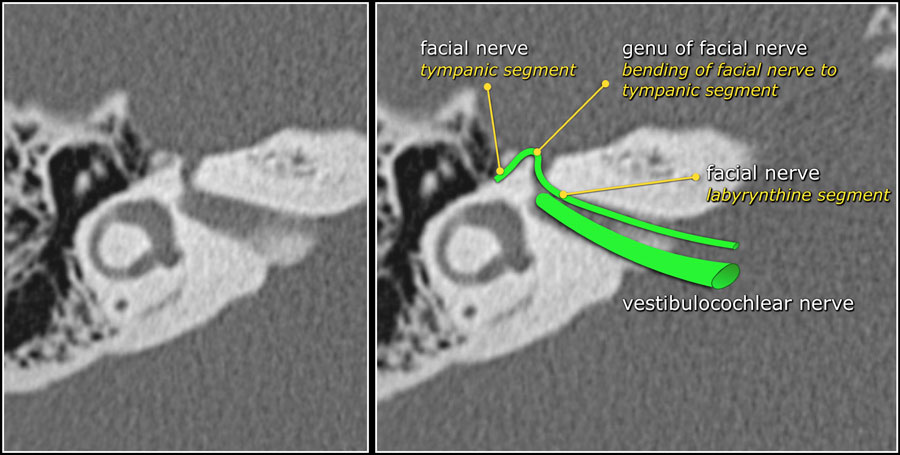
The Radiology Assistant : Temporal Bone - Anatomy 2.0
 radiologyassistant.nl
temporal bone anatomy nerve facial segment canal radiology petrous labyrinthine right coming axis angles geniculate auditory sharply nearly internal forward
CaseStacks.com - Chest CT Case #16
 www.casestacks.com
case casestacks
Exported LEFT TEMPORAL FIBROUS DYSPLASIA
 gamma.wustl.edu
temporal fibrous dysplasia ct bone submitted left fig
Exported left temporal fibrous dysplasia. Case casestacks. Temporal bone anatomy nerve facial segment canal radiology petrous labyrinthine right coming axis angles geniculate auditory sharply nearly internal forward