ct skull base anatomy
Frontal Sinus: Normal Anatomy & Variants. 9 Pictures about Frontal Sinus: Normal Anatomy & Variants : 3D reconstruction of COCHLEA | Radiology, Diagnostic imaging, Radiology, Superior View of the Skull Base | Neuroanatomy | The Neurosurgical and also Superior View of the Skull Base | Neuroanatomy | The Neurosurgical.
Frontal Sinus: Normal Anatomy & Variants
 uwmsk.org
frontal galli sinus crista variants coronal fracture bulla sinuses ethmoid normal recess anatomy ostium arrowheads pointing enlarged cells arrow note
3D Reconstruction Of COCHLEA | Radiology, Diagnostic Imaging, Radiology
 www.pinterest.com
jugular ct foramen anatomy radiology radiopaedia bone temporal scan head cochlea foramina carotid nerve pars ear internal artery nervosa 3d
Basics Of Ct Mri
 www.slideshare.net
temporal
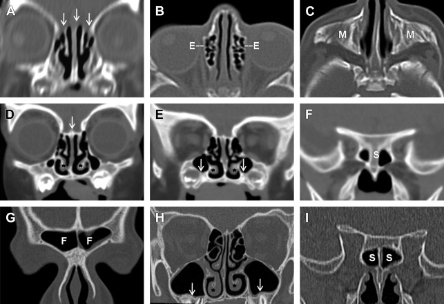
Normal Anatomy And Anatomic Variants Of The Paranasal Sinuses On
 radiologykey.com
paranasal tomography computed anatomic sinuses neuroimaging radiology
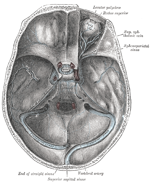
Transverse Sinuses - Wikidoc
 www.wikidoc.org
sinus sinuses transverse basilar cavernosus plexus anatomy skull cranial venous dural gray des base wikidoc foramen brain half right
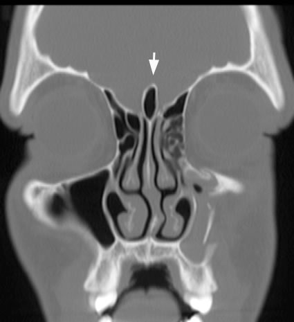
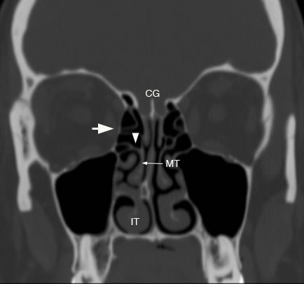
Ethmoid Sinus: Normal Anatomy & Variants
 uwmsk.org
cell ethmoid agger nasi basal sinus turbinate anatomy normal lateral ethmoidalis coronal lamellae middle lamina papyracea fovea cribriform plate inferior
Superior View Of The Skull Base | Neuroanatomy | The Neurosurgical
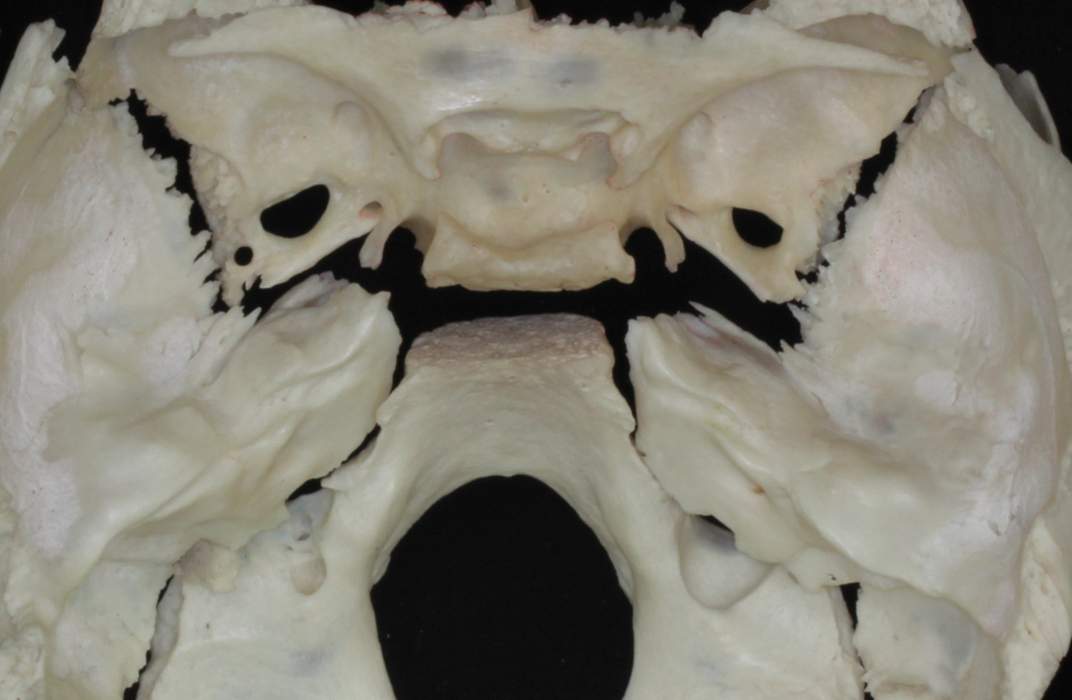 www.neurosurgicalatlas.com
correlation atlas neurosurgicalatlas
Lateral Lamella Of The Cribriform Plate: Software-Enabled Computed
 jamanetwork.com
cribriform plate lateral journals lamella neck clinical jamanetwork
Panoramic Superior Views Of Right And Left Orbits And Frontal, Ethmoid
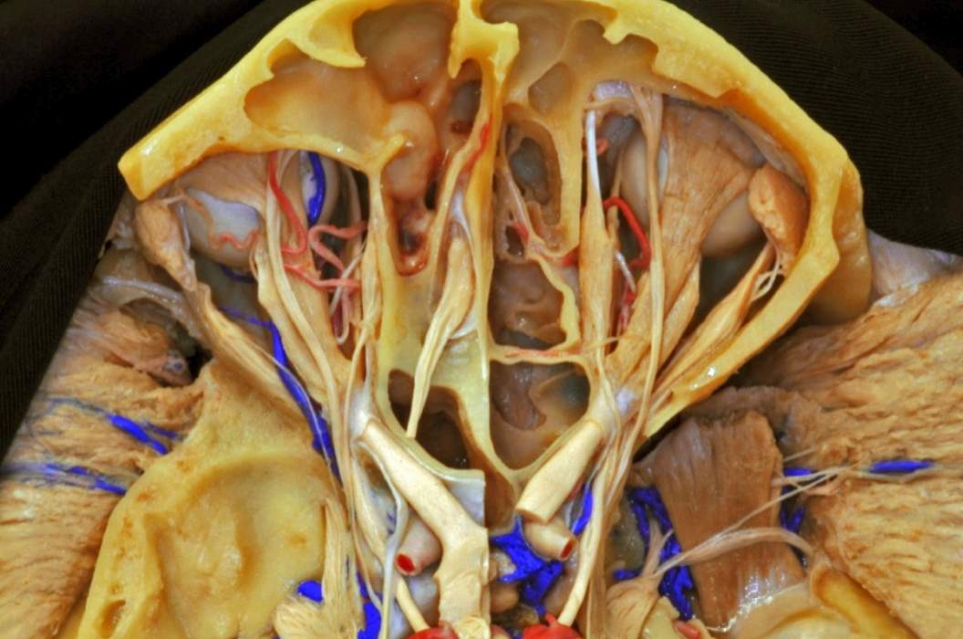 www.neurosurgicalatlas.com
superior panoramic left right ethmoid sphenoid views frontal sinuses surgical orbits atlas correlation neurosurgicalatlas
Jugular ct foramen anatomy radiology radiopaedia bone temporal scan head cochlea foramina carotid nerve pars ear internal artery nervosa 3d. Panoramic superior views of right and left orbits and frontal, ethmoid. Ethmoid sinus: normal anatomy & variants