ct pelvis anatomy
Urachal cyst: Clinical anatomy | Kenhub. 9 Images about Urachal cyst: Clinical anatomy | Kenhub : Pelvis computed tomograph (axial CT), Liver segments: annotated CT | Radiology Case | Radiopaedia.org | Liver and also Pelvis computed tomograph (axial CT).
Urachal Cyst: Clinical Anatomy | Kenhub
urachal cyst ct clinical anatomy axial kenhub case showing
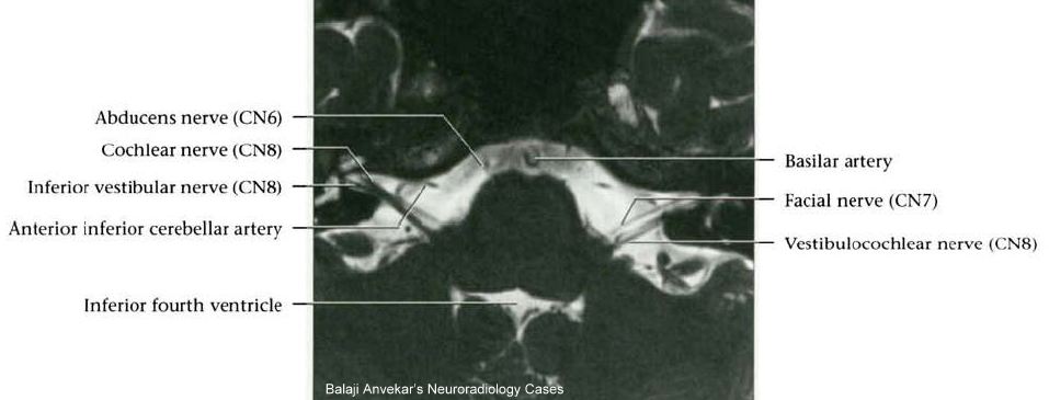
MRI Protocols: 7TH NERVE MRI Protocol(Facial Nerve)
 mriprotocol.blogspot.com
cranial nerves mri nerve imaging 7th anatomy facial protocol cn planning visualisation allowing effective provided examples such protocols
Anatomy Of The Pancreas
 www.aboutcancer.com
pancreas ct anatomy duct bile ercp
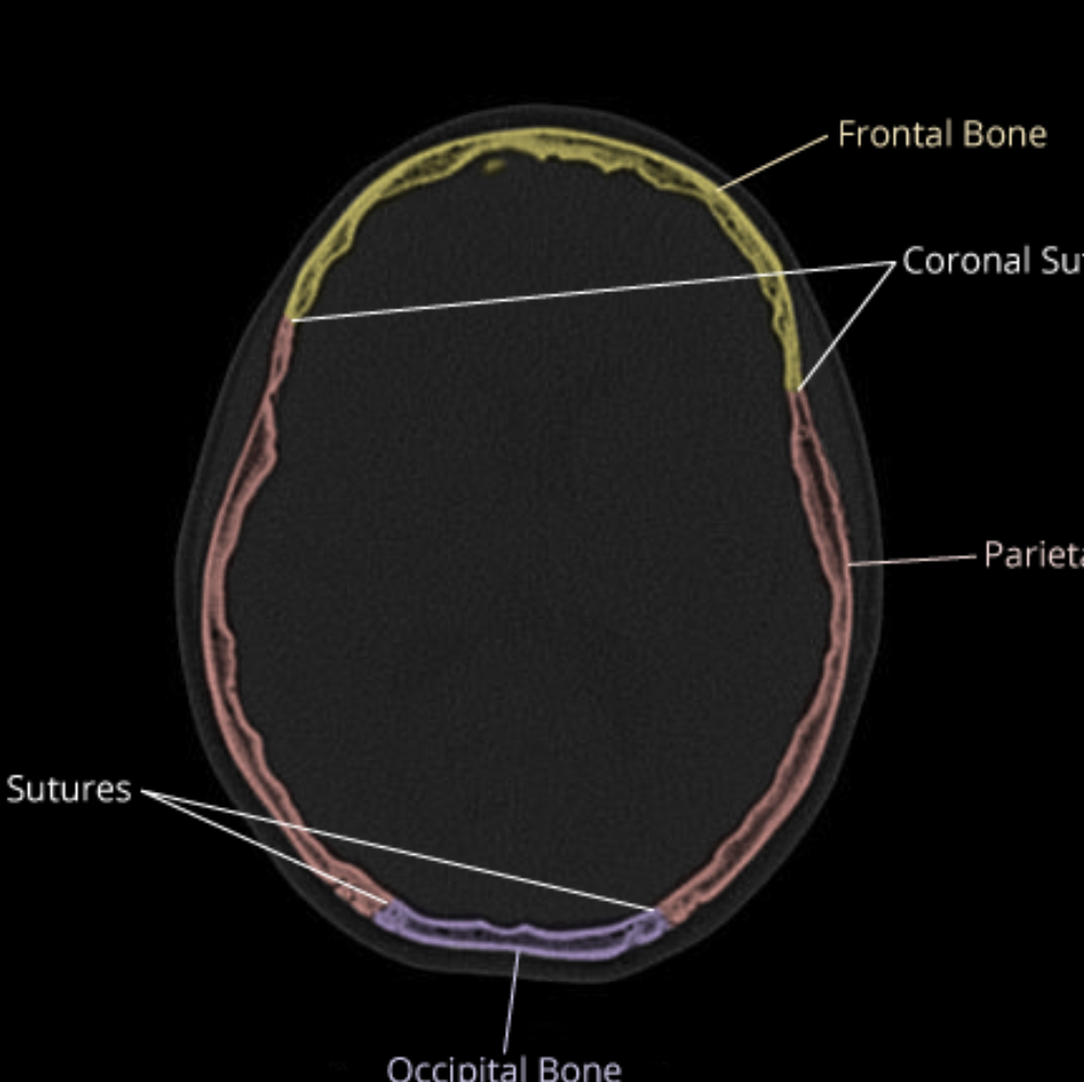
CaseStacks.com - Calvarial Anatomy On CT On Head CT
 www.casestacks.com
anatomy ct calvarial casestacks brain
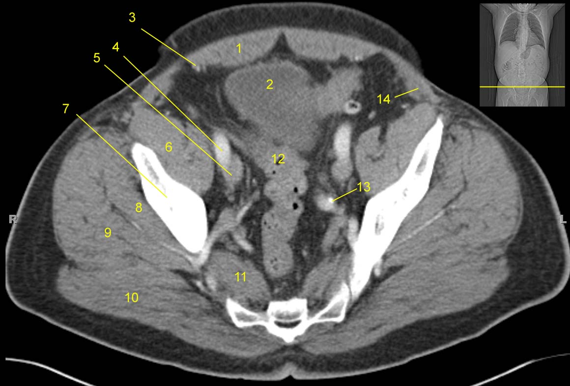
Pelvis Computed Tomograph (axial CT)
 jhurads4anatomy.com
ct pelvis anatomy scan pelvic muscle axial iliacus bone labeled computed cat tomograph legend study ss1 docs storage google
Ascites CT - Wikidoc
 www.wikidoc.org
ascites radiopaedia ct cholangiocarcinoma radiology case wikidoc fluid cases intraperitoneal venous phase portal due via
101 Spigelian Hernia | Radiology Key
 radiologykey.com
hernia spigelian ct abdominal anterior lateral radiology sigmoid semilunar radiologykey defect through
Liver Segments: Annotated CT | Radiology Case | Radiopaedia.org | Liver
 www.pinterest.co.kr
radiopaedia liver ct segments radiology annotated anatomy axial segmentation contrast ultrasound case radiological vascular nuclear medicine pelvis medical
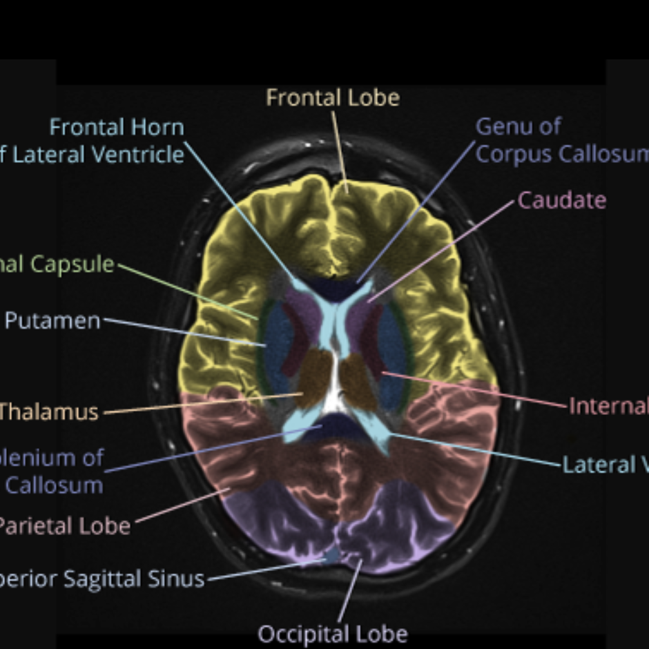
CaseStacks.com - MRI Brain Anatomy
 www.casestacks.com
brain mri anatomy casestacks
Ascites ct. Mri protocols: 7th nerve mri protocol(facial nerve). Pelvis computed tomograph (axial ct)